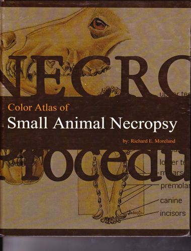
Color Atlas of Small Animal Necropsy
by Richard E. Moreland, Published 2009, FileType: PDF

Necropsy is the animal analogy to human autopsy. At its core, it is the systematic dissection and examination of an animal carcass to search for abnormal anatomical changes (lesions) in the tissues. It is generally used to determine the cause of death, but is also used to chronicle disease progression. Lavin’s Radiography for Veterinary Technicians, 5th Edition
Necropsy is the purest form of pathology. It involves the direct visualization of diseased organs and tissues (grossly and/ or microscopically) and can provide a wealth of information, not only about the animal being necropsied, but about the cause, progression, and possible outcome of diseases in other patients. Necropsy results can provide feedback on implemented therapies, and confirm or deny clinical assumptions and diagnoses.
Obviously, a knowledge of the normal anatomy is necessary to make a distinction between normal tissues and lesions. The proper, standardized necropsy procedures are designed to allow the prosecutor (the person doing the necropsy) maximal exposure of organs for maximum visualization of possible lesions.
Obtaining the maximum benefit and information from a necropsy requires not only knowledge of the proper necropsy dissection procedure, but knowledge of basic disease processes. In particular an understanding of basic pathology processes is paramount, starting with standard basic pathological definitions.
Direct Link For Paid Membership: –
This Book is Available For Premium Members Only (Register Here)
Unlock 3000+ Veterinary eBooks or Go To Free Download
Available for Free Membership: –
| Book Name: Color Atlas of Small Animal Necropsy | |
| Size : 91MB | Download Now : Click Here |
| Password : PDFLibrary.Net (If Required) | |
