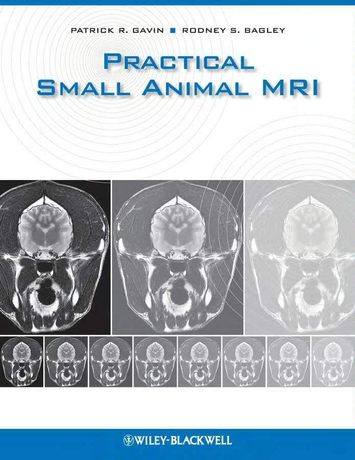
Practical Small Animal MRI is the seminal reference for clinicians using Magnetic Resonance Imaging in the diagnosis and treatment of veterinary patients.
Practical Small Animal MRI

Although MRI is used most frequently in the diagnosis of neurologic disorders, it also has significant application to other body systems. This book covers normal anatomy and specific clinical conditions of the nervous system, musculoskeletal system, abdomen, thorax, and head and neck. It also contains several chapters on disease of the brain and spine, including inflammatory, infectious, neoplastic, and vascular diseases, alongside congenital and degenerative disorders.
The superior visualization of soft tissues of the body has allowed for the imaging of virtually all disease processes. The improved conspicuity allows clinicians of multiple disciplines to have clear visualization of the disease process. This text is to provide examples of our experiences that have been gained over the past 21 years. In addition to our experiences, some of the examples come from my active collaboration with other sites. These include the IAMS Pet Imaging Centers in Vienna, Virginia, Raleigh, North Carolina, and Redwood City, California. Examples have also been used from Dr. Michael Broome’s Advanced Veterinary Medical Imaging Center in Tustin, California. These centers, coupled with the Washington State University cases make up the bulk of the material for the figures in the text, but other images come from Dr. Kelley Collins, Veterinary Imaging Center in Ambler, Pennsylvania, Oakridge Veterinary Imaging in Edmond, Oklahoma, Tacoma Veterinary MRI in Tacoma, Washington, and Veterinary Neurological Centers in Phoenix, Arizona.
The images chosen are realistic examples of common abnormalities. We have endeavored to provide good quality images, but not ones that cannot be readily obtained virtually all superconducting magnets. There are no permanent magnet images due to lack of availability of such images in our files. The images are shown with the patients right on the viewers left, unless otherwise indicated. The images are generally shown with the dorsal anatomic area to the top of the image, even when acquired differently. If the dependence of the image is important, that will be given in the figure legend.
Direct Link For Paid Membership: –
This Book is Available For Premium Members Only (Register Here)
Unlock 3000+ Veterinary eBooks or Go To Free Download
Direct Link For Free Membership: –
| File Size: | 25 MB | |
| Download Link: | Download | |
| Password: | PDFLibrary.Net (if Required) | |
