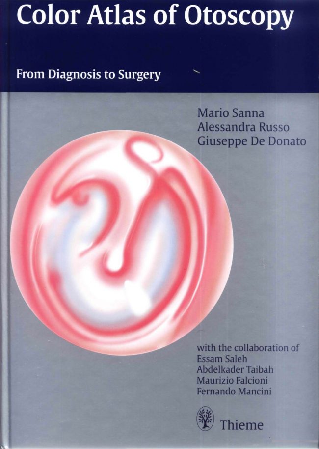
Color Atlas of Otoscopy PDF. Despite advances in diagnostic techniques and imaging modalities, otoscopy remains the cornerstone in the diagnosis of otologic diseases.
Color Atlas of Otoscopy PDF
Every otolaryngologist, pediatrician, or even general practitioner dealing with ear diseases should have a good knowledge of otoscopy. While otoscopy alone can establish the diagnosis in some cases, parameters such as history, or audiological and neuroradiological evaluation are required in others. An important aspect of this atlas is that it juxtaposes, when appropriate, the clinical picture, radiological diagnosis, and intraoperative findings with the otoscopic findings of the patient.
Needless to say, every patient should be considered as a whole and in some particular cases, the otoscopic findings might only be the “tip of the iceberg.” Otalgia, otorrhea, and granulations in the external auditory canal are manifestations of otitis externa, but when they persist, particularly in the elderly, they should arouse suspicion of malignancy.
Otitis media with effusion can be a simple disease when seen in children, whereas unilateral persistent otitis media with effusion in an adult may be the only sign of a nasopharyngeal carcinoma. A small attic perforation in the presence of facial nerve paralysis and sensorineural hearing loss may be all that is seen in a giant petrous bone cholesteatoma.
The manifestation of an aural polyp can vary from a mucosal polyp associated with chronic suppurative otitis media to the much less common but more dangerous glomus jugulare tumor. A small retro tympanic mass may represent an anomalous anatomy such as a high jugular bulb or an aberrant carotid artery. It may also represent frank pathology such as facial nerve neuroma, congenital cholesteatoma, or even enplaque meningioma.
In each chapter, a surgical summary that lists the different approaches for the management of the pathology dealt with is provided. Throughout the book, emphasis is on how the otoscopic view and the clinical picture may affect the choice of treatment and the surgical technique.
Direct Link For Paid Membership: –
This Book is Available For Premium Members Only (Register Here)
Unlock 3000+ Veterinary eBooks or Go To Free Download
Available for Free Membership: –
| Size : 10 | Download Now : Click Here |
| Password : PDFLibrary.Net (If Required) | |


This post contains broken links
link is nor working
Link Updated!