3D Joint Anatomy In Dogs, Main Joint Pathologies and Surgical Approaches
by
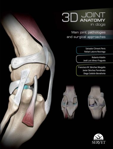
A visual guide with a strongly educational approach covering the main joints in the limbs of the dog. It shows the anatomical elements of each of these joints in three-dimensional diagrams. The views chosen for each case have been selected for a practical purpose, showing the position of the elements involved in the most commonly used surgical approaches. It also describes the key orthopaedic conditions affecting each joint and the most commonly used surgical approaches. It contains a large number of images and illustrations, and a selection of views presented in digital video format.
From the Publisher

3D Joint Anatomy in Dogs. Main joint pathologies and surgical approaches
This very instructive visual guide covers the main joints of the limbs in the dog by means of three-dimensional images. The main orthopedic problems of each joint and the most commonly used surgical approaches are also described. 3D Dog Anatomy Android App
This publication 3D Joint anatomy in dogs – Main joint pathologies and surgical approaches have three key noteworthy aspects.
Firstly, it is of doubtless clinical value, as it focuses on the six main joints of the dog’s body.
Secondly, it is developed in stages, from the clear and precise anatomic context of each and every joint, with a series of real images obtained by CT and MRI scans, x-rays and even fluoroscopes, and a collection of specially designed three-dimensional drawings, together with real plastinated sections to clarify and further doubts that the reader may have. Having provided a clear presentation of the anatomy, the book is complemented with a description of the most common pathologies in canine joints.

3D Joint Anatomy in Dogs. Main joint pathologies and surgical approaches
All this is required in order to perform the different orthopedic procedures required to treat these pathologies, brought together in a specific manner in this publication.
The project, which has brought together the work of anatomists, imaging specialists, and clinicians from a number of countries, was intended from the outset to be of direct application in orthopedics and use in daily clinical practice with dogs. The correlation of the CT and MRI images with real plastinated sections, which facilitate an understanding of the anatomical structures and the consequences of different pathologies, has therefore resulted in an entirely appropriate increase on the part of the authors of the number of images used in relation to the text.

3D Joint Anatomy in Dogs. Main joint pathologies and surgical approaches
Overall, and considering the importance of diagnostic imagining techniques, this work is not only of interest for clinical professionals but is also relevant to all the recent developments taking place in the field of cell therapy, regarding the regeneration of cartilage and control of inflammation, as the images provided through CT and MRI scans provide us with a clear and accurate picture of the evolution and monitoring of these aspects.
Given the above, we advise clinical staff to keep a copy of this project among their reference books, for clinical application and consultation purposes.
Direct Link For Paid Membership: –
