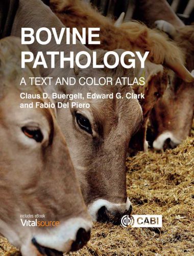
Bovine Pathology, A Text and Color Atlas by Claus D. Buergelt, Edward G. Clark, Fabio Del Piero May 2018. Illustrated with over 1,000 color images of the highest quality, Bovine Pathology: A Text and Color Atlas is a comprehensive single resource to identifying diseases in dairy cattle, feedlot cattle, and their calves. With summary text describing key features, the book correlates clinical information with pathology and differential diagnoses.
Bovine Pathology, A Text and Color Atlas

The text covers naked-eye macroscopic appearance through to microscopic pathology and the immunohistochemistry of infectious agents and tumor markers. Structured by major organ system, the disease entries follow a consistent format and clarity of display. This, combined with an integrated e-book, handy fact sheets, summary boxes and key points, helps aid understanding. Key features include:
– Over 1000 superb color images to illustrate the pathologies
– A thorough review of mainly western hemisphere diseases of cattle covering macroscopic appearance, microscopic appearance, and immunohistochemistry
– Synoptic layout, fact sheets, summary boxes, succinct legends and key bullet points support its use as a field guide or revision aid
– Organized by major organ system which ensures that vital facts can be found quickly
– A unique chapter covering calfhood diseases
Serving as an essential reference work for veterinary pathologists who perform bovine necropsies, veterinary residents and students, the book is also practical enough for bovine practitioners who need to investigate sudden death losses of cattle on the farm.
The book was designed with the needs of veterinary pathologists and practitioners in mind, and it should be a useful resource for the daily practice of bovine veterinarians, with emphasis on those working with feedlots. Veterinary students and veterinarians in training will also benefit greatly from the information contained in this book, which has examples of almost all lesions a bovine veterinarian should expect to see in his or her career. The book should prove a very valuable resource for veterinarians preparing for different specialty boards ….
A particularly useful feature of the atlas is the inclusion of brief text accompanying each image, which should prove of practical use when rapid consultation on the face of a particular case is required. An updated list of references after each chapter provides support for those readers who would like to dig deep into a particular condition. Images of immunohistochemistry and special stains add value to the discussion of each condition. Finally, and as a bonus, the book comes with a code that allows access to an electronic version of the book. This book is a must in any veterinary pathologist bookshelf and/or computer.
I found this book to be organized, thorough, visually appealing, and interesting. In it, there are things one sees every day and things one has rarely or never seen. From a Canadian perspective it is quite relevant, with a slight emphasis on feedlot and a western hemisphere bias. This book will definitely be useful to pathologists, students (who can still remember their histology), and bovine practitioners (who probably can’t), and might even provide some incentive to do more postmortems in the field to find some of these interesting lesions.
Direct Link For Paid Membership: –
This Book is Available For Premium Members Only (Register Here)
Unlock 3000+ Veterinary eBooks or Go To Free Download
Available for Free Membership: –
| Size : 6 MB | Download Now : Click Here |
| Password : PDFLibrary.Net (If Required) | |

1
5