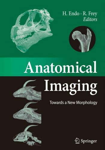
Anatomical Imaging: Towards a New Morphology
By H. Endo, H. Endo, R. Frey, Year 2008, File Type: PDF

This book presents selected works of contemporary evolutionary morphologists and includes such topics as broad scale reconstructions of the brain and ear of dinosaurs, inference of locomotor habits from cancellous bone architecture in fossil primates, and a comparison of the independently evolved manipulating apparatuses in the lesser and giant pandas. Insight is provided into the application of modern noninvasive technologies, including digital imaging techniques and virtual 3D reconstruction, to the investigation of complex anatomical features and coherences. In combination with traditional methods, this allows for the formulation of improved hypotheses on coordinated function and evolution. The creation of virtual translucent specimens makes it possible to realize the age-old dream of the classical anatomists: looking through the skin into the inner organization of an organism. On full display here is the dramatic and promising impact that modern imaging techniques have on scientific progress in evolutionary morphology.
From early on, anatomical imaging proceeded along three tracks that promoted each other mutually. One track consisted of human beings interested in the organic structure of animals and humans. This track comprises Paleo- and Neolithic hunters, shamans and clairvoyants of Sumeran and Babylonian times, dissecting artists of the Middle Ages, e.g., Leonardo da Vinci, and leads us to skilled and creative scientific anatomists of modern times.
The second one consisted of technologies involved in the processing of images. Thus, a short walk through the history of anatomical imaging will bring us from cave paintings via written information only, via drawings, paintings, engravings, lithographies and prints to black and white photography, roentgenography, analogue colour photography, digital photography, offset printing, analogue and digital video, ultrasound imaging, magnetic resonance imaging, computed tomographic scanning and software-based three-dimensional reconstructions. Veterinary Anatomy Coloring Book
The third one consisted of the media available or specifically made as a carrier for the images. It started with rock surfaces, then clay tablets, continued with parchment and paper, wooden blocks, copper plates, lime stones and brought us to photographic film, roentgen film and computer screens.
