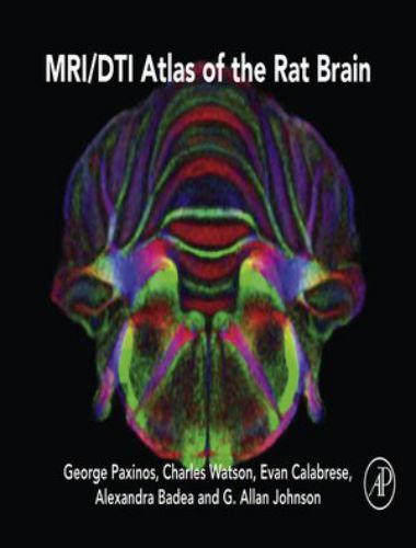
MRI/DTI Atlas of the Rat Brain
by George Paxinos, Charles Watson, Evan Calabrese, Alexandra Badea, Year 2015, File Type: PDF

MRI/DTI Atlas of the Rat Brain addresses the MRI/DTI resolution/contrast obtained at the Duke Center for In Vivo Microscopy, which has surpassed that of any other lab by nearly 400x, with images that are satisfactory for the identification of 80%+ of structures previously labeled in Rat Brain in Stereotaxic Coordinates.
This new atlas, from the best imaging/cartography team working in neuroscience today, fully complements the work in Rat Brain in Stereotaxic Coordinates, and will become a new landmark contribution to neuroanatomy as the first and only truly reliable MRI/DTI atlas of the rat brain. Atlas of Equine Ultrasonography
- Ninety-six coronal levels from the olfactory bulb to the pyramidal decussation are depicted
- Delineations primarily made on the basis of direct observations on the MRI contrasts
- Each of the 96 open book pages displays four items— top left the directionally colored fractional anisotropy image derived from DTI (DTI – FAC), top right the diffusion-weighted image (DWI), bottom left the gradient recalled echo (GRE), and bottom right the diagram. The diagram is the synthesis of the information derived from these three images and the two additional images, which we do not display (ARDC and RD). This is repeated for 96 coronal levels, which makes the levels 250 μm apart.
- The FAC images are shown in full color
- The orientation of sections corresponds to that in Paxinos and Watson’s The Rat Brain in Stereotaxic Coordinates (2014)
- The images have been obtained from 3D isotropic population averages (number of rats=5). All abbreviations of structure names are identical to the Paxinos & Watson histologic atlas
The first atlas to offer comprehensive MRI and DTI coverage of the rat brain, from the most successful team of cartographers in neuroscience today.

5