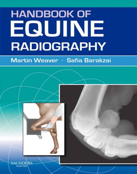
The Handbook of Equine Radiography 1st Edition is a practical and accessible “how-to guide to obtaining high-quality radiographs of the horse.
Handbook of Equine Radiography, 1st Edition

It covers all aspects of taking radiographs of the commonly examined regions (lower limbs and skull) as well as less frequently examined areas (upper limbs, trunk).
The main part of the book consists of diagrams to illustrate the positioning of the horse and the radiography equipment. For each view a benchmark example of a normal radiograph is illustrated. The accompanying text for each radiographic view succinctly presents the most relevant aspects.
Practically orientated, and including chapters covering such key areas as radiation safety in equine radiography and patient preparation, plus a trouble-shooting section, the Handbook of Equine Radiography is an indispensable guide to practitioners in all countries engaged in equine work.Clear diagrams illustrate the positioning of the horse and the radiography equipment Contains all the information required to radiograph a horse Accessible to veterinary surgeons who obtain most of their radiographs in the field.
All radiographic images reflect a pattern of x-ray photons (particles of energy) that have passed through the subject with a proportion being absorbed, depending on the nature of the tissue (fluid, soft tissue or bone). X-rays are a form of energy, which are emitted from tungsten when the metal is subjected to a stream of electrons. In traditional radiography using non-screen film the x-rays emitted from the x-ray machine pass through the patient, subsequently causing ionisation of silver halide embedded in the emulsion coating the film (exposure). The introduction of fluorescent intensifying screens into radiographic cassettes adds one further step to the process: the x-rays cause light to be emitted from the screens and it is this light that ionises the silver. Because one x-ray photon produces many light photons the x-ray machine exposure settings can be reduced when screens are used. The latent image present on the film is then converted to the familiar radiographic image by processing the film through liquid developing and fixation chemicals.
Password: pdflibrary.net
