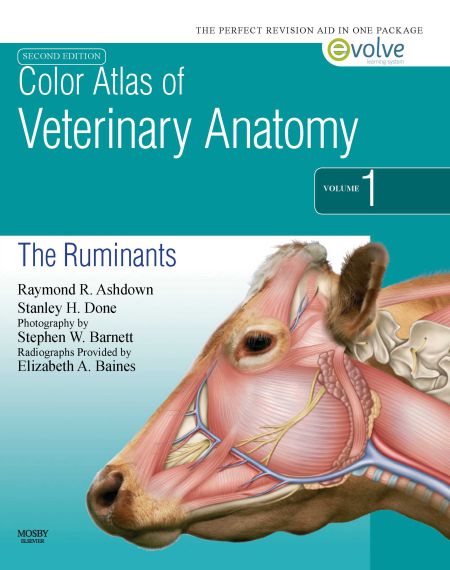
Color Atlas of Veterinary Anatomy, The Ruminants, 2nd Edition by Raymond Ashdown Stanley Done Stephen Barnett was published 13th February 2010.
Color Atlas of Veterinary Anatomy, The Ruminants, 2nd Edition

This book is intended for veterinary students and practising veterinary surgeons. Important features of topographical anatomy are presented in a series of full-colour photographs of detailed dissections. The structures are identified in accompanying coloured line drawings, and the nomenclature is based on that of the Nomina Anatomica Veterinaria (1992); latin terms are used for muscles, arteries, veins, lymphatics and nerves, but anglicized terms are used for all other structures. When necessary, information needed for interpretation of the photographs is given in the captions. Each section begins with photographs of regional surface features taken before dissection, and complementary photographs of an articulated bovine skeleton illustrate the important palpable bony features of these regions. All dissections and photographs have been specially prepared for this book.
The cattle (two cows and four calves) were all Jerseys and the three goats were of the British Saanen breed. The specimens were embalmed, for the most part, in the standing position using methods routinely employed in the Department of Anatomy at the Royal Veterinary College. Every effort was made to ensure that the final position corresponded to that of normal level standing. In most cases red neoprene latex was injected into the arteries. The dissections follow the pattern of prosections used for teaching at the Royal Veterinary College for many years. The photographs of the adult bulls were taken at the Milk Marketing Board’s cattle breeding center at Bletchley.
The aim of these dissections and photographs is to reveal the topography of the animal as it would be presented to the veterinary surgeon during a routine clinical examination. Therefore, lateral views pre-dominate and we have, as far as possible, avoided photographs of parts removed from the body or the use of views from unusual angles, or of unusual bodily positions. It is our earnest hope that this book will enable students and veterinary surgeons to see, beneath the outer surface of the animals entrusted to their care, the muscles, bones, vessels, nerves and viscera that go to make up each region of the body and each organ system.
Password: pdflibrary.net
