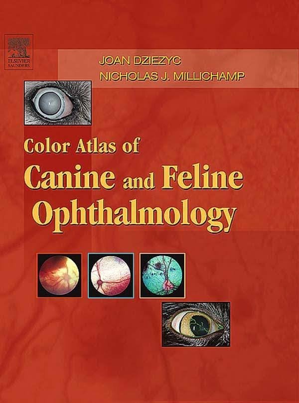
Color Atlas of Canine and Feline Ophthalmology PDF. When initially approached by Saunders to produce an atlas of canine and feline eye diseases, we hesitated. However, over the years we had accumulated many photographs that we knew would be a great source of material for the atlas.
Color Atlas of Canine and Feline Ophthalmology PDF
“How difficult can it be to produce an atlas?” we asked ourselves. Well … the daunting task soon became readily apparent! Sorting through more than 20 years’ worth of photographs, selecting 10,000 of those images to scan electronically, and then distilling that number down to the 1000 or so we felt best represented the conditions we would be presenting was more difficult than we could have imagined.
With this final set of images, we now have developed a new atlas that depicts normal and diseased eyes as they might be seen during an ocular examination by any general practitioner. We have also included some material that would be of benefit to those veterinarians with a more specialized interest in ophthalmology and to residents in ophthalmology. Furthermore, there are many fundus photographs, representing both normal and abnormal views, which are not available in most other atlases.
The aim of this atlas is simply to illustrate the canine and feline eye and its lesions. There are excellent ophthalmology references and textbooks available, and we do not intend to duplicate what is written elsewhere. In fact, the text in this atlas is intentionally limited to a brief introduction at the beginning of each section and legends that describe simply the structure or condition presented. We do not provide detailed information on etiology, diagnosis, and treatment.
In ophthalmology, more than any other specialty, the clinician relies heavily on what he or she can actually see. Visual recognition of structures and lesions is key to making a proper diagnosis. We believe the strength of this atlas lies in its comprehensive depiction of the many normal and abnormal features of the eye that will be encountered during an ocular examination.
Proper identification of what is seen by the clinician’s own eye is the most critical step of the process when treating an ocular problem. The goal of this atlas is to provide as much assistance as possible toward that end. For more thorough understanding of the anatomy and pathophysiology of the eye, we refer the reader to a more general ophthalmology textbook. Following the conventional format of most of those books, the chapters and illustrations in this book are arranged from anterior to posterior and include the orbit. Throughout the atlas there are numerous images of normal ocular features as well as lesions commonly seen in practice. We have also included a number of illustrations of conditions that less frequently occur.
Direct Link For Paid Membership: –
This Book is Available For Premium Members Only (Register Here)
Unlock 3000+ Veterinary eBooks or Go To Free Download
Direct Link For Free Membership: –
| Book Name: | Color Atlas of Canine and Feline Ophthalmology PDF | |
| File Size: | 63 MB | |
| File Format: | ||
| Download Link: | Click Here | |
| Password: | PDFLibrary.Net (if Required) | |

