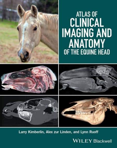
Atlas of Clinical Imaging and Anatomy of the Equine Head presents a clear and complete view of the complex anatomy of the equine head using cross-sectional imaging.
Atlas of Clinical Imaging and Anatomy of the Equine Head

Provides a comprehensive comparative atlas to structures of the equine head Pairs gross anatomy with radiographs, CT, and MRI images Presents an image-based reference for understanding anatomy and pathology Covers radiography, computed tomography, and magnetic resonance imaging.
Atlas of Clinical Imaging and Anatomy of the Equine Head presents a clear and complete view of the complex anatomy of the equine head using cross-sectional imaging.
- Provides a comprehensive comparative atlas to structures of the equine head
- Pairs gross anatomy with radiographs, CT, and MRI images
- Presents an image-based reference for understanding anatomy and pathology
- Covers radiography, computed tomography, and magnetic resonance imaging
Atlas of Clinical Imaging and Anatomy of the Equine Head presents a clear and complete view of the complex anatomy of the equine head using cross-sectional imaging. The gross anatomy of a one-centimeter section of the equine head is compared to identical slices in CT and MRI in the transverse, sagittal, and dorsal planes. To aid in the identification of clinically important structures, the book covers oral, dental, nasal, sinus, ophthalmic, auricular, laryngeal, hyoid apparatus and tongue structures.
The atlas offers more than 300 gross photographs, radiographs, CT images, and MRI images, with all structures indicated using color-coded labels. Veterinary students, equine practitioners, surgeons and imaging specialists who wish to foster a clear understanding of the anatomy of the structures involved in the equine head will find Atlas of Clinical Imaging and Anatomy of the Equine Head an essential resource.
RAR File Password: pdflibrary.net
Password: pdflibrary.net
