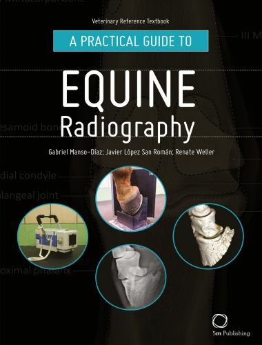
A Practical Guide to Equine Radiography is designed to accompany the clinical veterinarian either within a hospital setting or out in the field. The handbook offers an informative step-by-step guide to obtaining high quality radiographs, consistently.
A Practical Guide to Equine Radiography

Each chapter focuses on a separate region of the horse, offering tailored material in a clear and concise way – suitable for accessing as use of a reference. This manual offers a comprehensive guide to taking radiographs by including: clinical indications for the radiographic area of interest; equipment required; preparation and setup, with photographs; projections suitable for the radiographic area of interest, with photographs; example x-ray with labels; and three-dimensional image to demonstrate normal anatomy. This book A Practical Guide to Equine Radiography is an essential tool for all practicing equine veterinarians and students alike.
Features provided in the book will guide the veterinarian through the stages of taking and interpreting normal radiographs and include:
- Clinical indications of radiographic areas of interest in the horse
- Equipment required
- Preparation and setup guides, supported by photographs
- Projections focusing on radiographic areas of interest, aided by photographs
- x-rays presented with detailed labels, providing a close-up view of skeletal structures
- Three dimensional images demonstrating normal anatomy
A Practical Guide to Equine Radiography is an essential tool for equine practitioners, veterinary students and para-professionals.
| Get Hard Copy | PDF Download |
Password: pdflibrary.net
